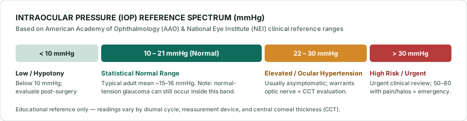
Complete Eye Pressure (IOP) Reference Chart
Intraocular pressure is measured in millimeters of mercury (mmHg)—the same pressure unit used for blood pressure, though the two systems are independent. Because each person's optic nerve tolerates pressure differently, ophthalmologists interpret your number along a clinical spectrum rather than a single pass/fail line:
| IOP Range (mmHg) | Clinical Classification | Typical Next Step |
|---|---|---|
| Below 10 mmHg | Low Eye Pressure (Hypotony) | Clinical review, especially after eye surgery, trauma, or uveitis. |
| 10 – 21 mmHg | Statistical Normal Range | Routine dilated eye exams; still evaluate optic nerve for normal-tension glaucoma. |
| 22 – 24 mmHg | Borderline / Mild Elevation | Confirm with Goldmann applanation, check corneal thickness (CCT), baseline OCT/visual field. |
| 25 – 30 mmHg | Moderate Ocular Hypertension | Prompt ophthalmologist evaluation; discuss risk profile and possible pressure-lowering drops. |
| Above 30 mmHg | High Risk Elevation | Urgent clinical assessment; high risk of progressive optic nerve injury if sustained. |
| 50 – 80 mmHg + Pain | Acute Angle-Closure Crisis | Immediate Emergency Room / Ophthalmic ER visit—can cause irreversible vision loss within hours. |
Normal Eye Pressure by Age Group
Unlike blood pressure targets that shift sharply across adulthood, the standard 10 to 21 mmHg adult IOP reference band remains largely consistent from young adulthood through senior years. Key age-related clinical nuances include:
- Infants and young children: Baseline IOP is naturally lower—often 8 to 15 mmHg—and gradually rises toward adult values during adolescence. Pediatric measurements often use drop-free rebound devices like the iCare ic100 or ic200 to avoid anxiety-induced squeezing that artificially spikes readings.
- Adults aged 20–40: Population averages hover near 14–16 mmHg. Routine comprehensive eye exams establish a personal baseline before age-related drainage resistance develops.
- Adults aged 40–60+: While the statistical cutoff stays 21 mmHg, age is an independent risk factor for primary open-angle glaucoma. The American Academy of Ophthalmology recommends baseline comprehensive eye evaluations at age 40, with closer intervals for individuals with a family history of glaucoma, African or Hispanic ancestry, high myopia, or diabetes.
Why "Normal" Pressure Is Not Always "Safe": Two Critical Exceptions
1. Ocular Hypertension (High IOP, Healthy Nerve)
Ocular hypertension means eye pressure consistently measures above 21 mmHg, yet optic nerve imaging (OCT), dilated examination, and visual field testing show zero glaucomatous damage. Landmark findings from the NIH-sponsored Ocular Hypertension Treatment Study (OHTS) demonstrated that not every patient with ocular hypertension converts to glaucoma. Doctors weigh four major risk factors—higher baseline IOP, older age, thinner central corneal thickness (CCT), and larger optic nerve cup-to-disc ratio—when deciding whether to monitor closely or start preventive eye drops.
2. Normal-Tension Glaucoma (Normal IOP, Nerve Damage)
Conversely, a substantial subset of glaucoma patients develop progressive optic nerve thinning and blind spots even though their office IOP never exceeds 21 mmHg. In normal-tension glaucoma, the optic nerve is unusually vulnerable to moderate pressure or reduced ocular blood flow. Treatment still focuses on lowering IOP further (often targeting a 25–30% reduction below the patient's untreated baseline).
Five Factors That Make Your Eye Pressure Reading Fluctuate
- Diurnal (24-hour) rhythm: Healthy eyes commonly vary by 2 to 6 mmHg over 24 hours, while glaucomatous eyes can swing 10 mmHg or more. Peaks frequently occur overnight or shortly after waking.
- Body posture: Lying flat (supine) increases episcleral venous pressure and raises IOP by several mmHg compared with sitting upright.
- Central Corneal Thickness (CCT): Goldmann applanation tonometry assumes an average corneal thickness near 520–545 µm. Thick corneas (>575 µm) resist flattening and read falsely high; thin corneas (<520 µm) flex easily and read falsely low. Test how corneal thickness shifts published estimates with our IOP & CCT Reference Calculator.
- Measurement device: Quick screening tools like air-puff (non-contact) tonometers can read slightly differently from slit-lamp Goldmann applanation. See our Tonometer Types Guide for why doctors confirm elevated screenings with contact applanation.
- Exam technique: Holding your breath, squeezing your eyelids tightly, wearing a tight collar or necktie, or drinking large volumes of water rapidly just before an exam can temporarily elevate readings.
Emergency Red Flags: When High Eye Pressure Cannot Wait
Chronic elevated eye pressure is painless, which is why regular exams matter. Acute angle-closure glaucoma is completely different: the eye's drainage angle blocks suddenly, driving pressure to 50–80 mmHg within hours. Go to an emergency department immediately if you or someone else experiences:
- Sudden, severe, throbbing pain in one eye or around the brow
- Abruptly blurred or foggy vision
- Seeing rainbow-colored halos or rings around lights
- A visibly red eye with a fixed, mid-dilated pupil
- Intense headache accompanied by nausea or vomiting
Clinical Source Verification: Reference values and emergency criteria on this page align with published clinical guidance from the American Academy of Ophthalmology (AAO) and the National Eye Institute (NEI, National Institutes of Health). Always consult a licensed ophthalmologist or optometrist to establish your individual target IOP.